facebook
Esophageal Cancer Treatment in Navi Mumbai

Esophageal Cancer

Esophagus is the upper part of the gastrointestinal tract that connects the mouth to the stomach. Your esophagus is not fully equipped to handle stomach acids, so when the stomach acids constantly regurgitate to the esophagus, its lining gets damaged, which creates a series of destructive changes leading to Barrett's esophagus and esophageal cancers.

There are two main types of esophageal cancers - Squamous cell carcinoma and adenocarcinoma.

  • Squamous cell carcinoma
    It is a cancer of the squamous cells that line the inner esophagus and is more likely to occur in the upper and mid portions of the esophagus. Cancer often begins as a plaque or a small outgrowth of cells that ultimately grows to form cancer.
  • Adenocarcinoma
    affects the lower one-third of the esophagus and is characterised by the transformation of squamous cells of the esophagus into gland cells. Many cases of adenocarcinoma are caused mainly by Barett's esophagus. Barett's esophagus is a condition that arises from repeated exposure of the esophagus to the stomach acids.

    Esophageal cancer is more likely to occur in male patients. The male: female ratio is 3:1. The onset is later in life, 55-60 years. Esophageal cancer is one of the most aggressive cancers, and the prognosis is usually bad.

Squamous cell carcinoma

  • Smoking
  • Alcohol consumption
  • Infection with human papillomavirus (HPV)
  • Diet low in fruits and vegetables
  • Drinking hot liquids
  • Atrophic gastritis
  • Poor oral hygiene

Adenocarcinoma

  • Gastroesophageal reflux disease (GERD) Gastroesophageal reflux disease refers to a condition where stomach acid flows back into the esophagus. The presence of various acids in the esophagus can lead to esophageal erosion, Barett's esophagus, and several other complications. Barrett's esophagus has invasive potential, that is, the potential to change into cancer, most particularly adenocarcinomas.
  • Smoking and alcohol
  • Obesity Because of increased fat in the visceral organs, the esophageal sphincter undergoes increased pressure, increasing the incidence of GERD in such patients
  • H. pylori infection
  • Cholecystectomy

About half of the patients with esophageal cancers do not report any symptoms. Therefore, it is very important to undergo periodic medical checkups to find out cancers at an early stage, when it has not progressed to a point where it is incurable.

  • Progressive dysphagia: from solid foods to solids and liquids
  • Odynophagia or pain with swallowing
  • Regurgitation
  • Aspiration
  • Reflux (if there is concomitant GERD)
  • Hematemesis
  • Malena
  • Anemia
  • Constitutional symptoms
  • Fatigue
  • Night sweats
  • Weight loss
  • Other signs and symptoms
  • Dry cough: may be caused by aspiration and/or aspiration pneumonia
  • Hoarseness of voice
  • Stage 0
    Cells with abnormal morphology, not necessarily malignant, are found in superficial layers of cells that line the esophagus
  • Stage 1
    The cancerous cells are confined to the lining of the esophagus
  • Stage 2
    Cancer penetrates the deeper walls of the esophagus and spreads to a few nearby lymph nodes
  • Stage 3
    The cancerous cells invade the deep inner muscular layer of the esophagus. It may even invade more lymph nodes and involve nearby organs
  • Stage 4
    Cancer has metastasised to the distant organs or distant lymph nodes. It is the most advanced stage and has a very poor prognosis.

The diagnosis of esophageal cancer starts with a review of your symptoms and medical history and carrying out a thorough physical examination by your doctor. To save time and your financial resources, the investigations are carried out in a specific order.

  • Barium swallow
    You are given a drink that covers your esophagus. When visualised on an X-Ray, this drink highlights any abnormalities in the esophagus and helps your doctor locate those.
  • Endoscopic ultrasound
    A thin, lighted tube called an endoscope is passed down through the throat to the esophagus, which uses ultrasonic waves to provide the doctor with information regarding the extent of the local invasion of cancer.
  • Endoscopy with biopsy
    The doctor takes a small portion of the suspected tissues during endoscopy and sends them for histopathological examination.
  • To assess for metastasis
    CT scan is done to find out if cancer has metastasized to the lungs or liver. If cancer is not seen in these locations, a PET scan is ordered. A PET scan is more efficient than a CT scan and can sometimes locate cancers missed by a CT scan. We can sometimes go directly for a PET CT scan which combines the technologies of both the PET scan and CT scan. If these tests locate cancer outside the esophagus, the standard protocol is to conduct a biopsy of the tissue.

It is important to know how far the malignant cells have penetrated into the esophagus and where it is located to treat cancer appropriately. In the beginning, the malignant cells usually invade only the superficial layers of the esophagus and, therefore, are easier to cure. As it advances, the cancerous cells start penetrating the deeper layers of the esophageal wall. It may even invade the lymph nodes and other organs with further progression. When cancer is limited to the superficial stage, it is called the early stage of the disease. When it reaches the deeper layers of the esophagus or reaches the lymph nodes, it is called a locally advanced disease. When it reaches the distant organs, it is said to be in later stages. There are various modalities used to treat cancer of the esophagus, including esophagectomy, endoscopic mucosal resection, photodynamic therapy, chemotherapy, and radiation. These can sometimes be used in combination. The preferred treatment option depends upon the stage of cancer and the depth of its penetration.

  • If superficial, limited to mucosa
    Endoscopic resection can treat cancer. No surgery is needed.
  • Lesions penetrating the sub-mucosa
    Surgical resection ( esophagectomy) with lymphadenectomy (negative lymph nodes) is considered.
  • Lesions invading muscularis propria with affected lymph nodes
    Surgical resection with neoadjuvant chemotherapy.
  • Metastatic disease
    Palliative treatment, esophageal stent, chemotherapy, and radiotherapy.

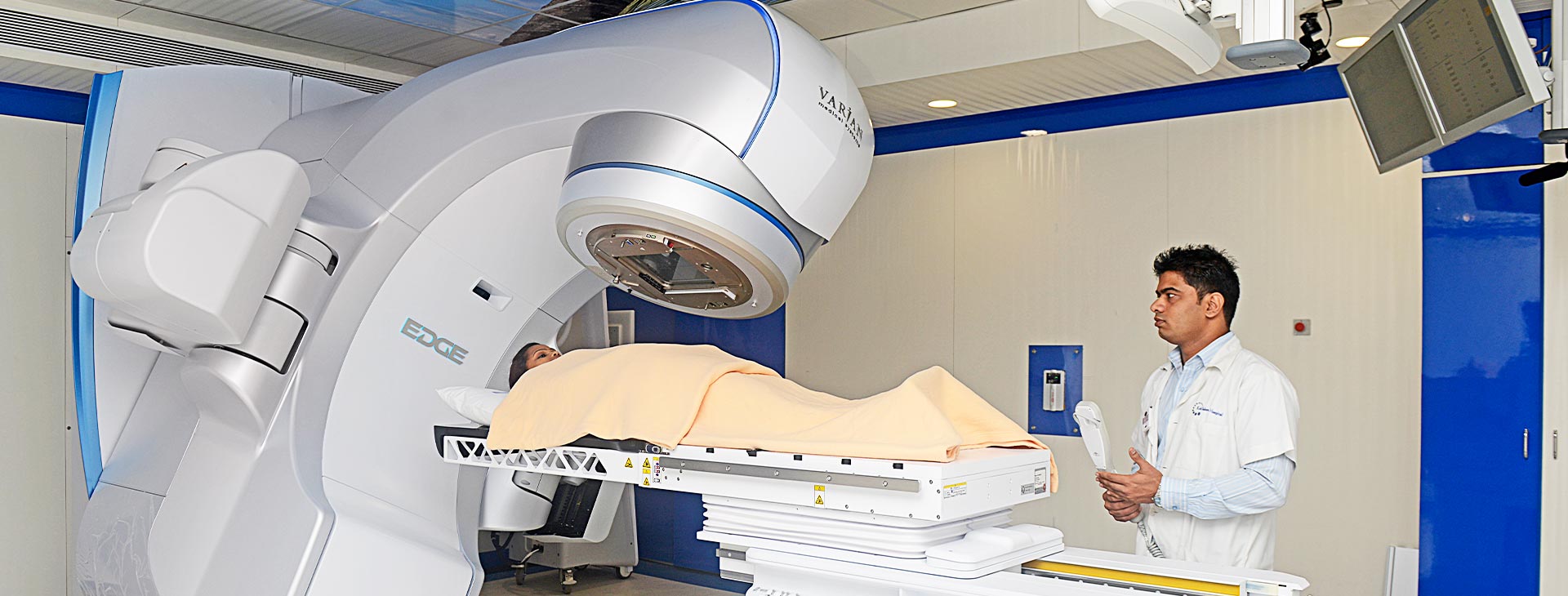
Kokilaben Dhirubhai Ambani Hospital, Navi Mumbai specializes in the treatment of many cancers. Our healthcare team discusses the treatment options with its risk and benefits to the patient's family and together curates the best plan for his condition. The surgeries testing esophageal cancers are quite challenging to perform and demand a multidisciplinary approach and expertise and precision by the surgeon. They can only be done in a big, well-equipped hospital-like Kokilaben Dhirubhai Ambani Hospital, Navi Mumbai with a multidisciplinary team proficient of addressing all the problems that the surgery can manifest and are comfortable dealing with any potential complications.