The Department of Interventional Radiology at Kokilaben Dhirubhai Ambani Hospital, Navi Mumbai involves conducting minimally invasive image-guided techniques and procedures employed for the diagnosis and treatment of disorders of the head, neck, and spinal cord.
Our goal is to offer optimum minimally invasive diagnostic and therapeutic services for a diverse range of conditions and disorders involving the head and neck area, brain, and spinal cord. Our team comprises dedicated medical experts who have extensive experience in delivering neuroradiological treatment and embolisation services for arteriovenous malformations (AVM), intracranial aneurysms, and dural arteriovenous fistulas (DAVF). Additionally, we offer treatment and management of carotid artery stenosis, stroke, cavernomas, subarachnoid haemorrhage (SAH), developmental venous abnormalities (DVA), spinal vascular malformations, superficial vascular malformations, and many more conditions.
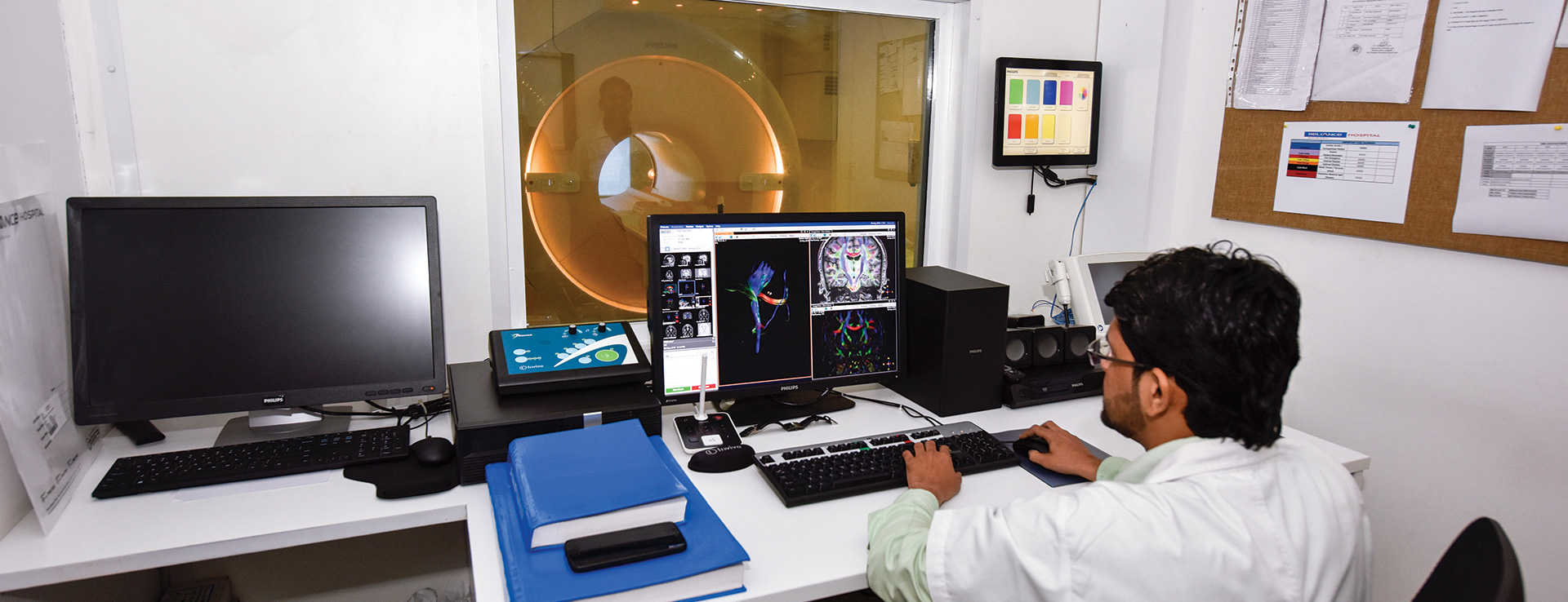
Our hospital is equipped with state-of-the-art infrastructure that our endovascular and interventional specialists use to deliver therapeutic agents percutaneously. The benefits of these procedures include shorter hospital stays and reduced recovery periods. Apart from these, the complications associated with these are minimal. Our experts are also trained in spinal angiography and diagnostic cerebral and head and neck angiography.
Our team works in close collaboration with experts from various other departments of Kokilaben Dhirubhai Ambani Hospital, Navi Mumbai so that multidisciplinary treatment and care are provided to the patients. These departments include the Department of Vascular Surgery, Department of Anaesthesiology, Department of Ophthalmology, Bone and Joint Centre, and the Department of Plastic Surgery. Depending on the type of patient, the experts from the respective departments are consulted to prepare tailored treatment plans.
Cerebral angiography is an interventional procedure that involves using contrast material and X-rays to determine how well the blood is flowing in the arteries of the brain.
Not everyone with arterial blockages requires cerebral angiography. It is usually recommended only if the doctor wants to have more information to formulate the treatment plan. This is because it is an invasive procedure and poses some risks, so it is avoided unless it is necessary to do it.
Sometimes, angiography can be used for therapeutic purposes, that is, for treating certain conditions involving the blood vessels of the brain and the neck.
Cerebral angiography is used for the diagnosis of certain conditions that involve the blood vessels of the brain and the neck, such as arteriosclerosis, aneurysms, vasculitis or inflammation of the blood vessels, arteriovenous malformations, blood clots in the vessels, brain tumours, and tears in the lining of an artery. It also helps doctors determine the causes of certain symptoms, such as severe headaches, slurred speech, memory loss, dizziness, blurred vision, weakness or numbness, and loss of balance or coordination.
Sometimes, angiography is used to help treat some of the conditions involving the blood vessels of the neck and brain.
During the procedure, the patient is asked to lie down on an X-ray table. The head is held with the help of a tape, strap, or sandbag so that it stays stationary during the procedure. A mild sedative is given for relaxation, and ECG leads are placed on the arms and the legs so that cardiac activity can be monitored throughout. After this, the groin area is cleaned and numbed with local anaesthesia. A catheter is introduced through an incision into the artery. It is carefully moved through the blood vessels of the abdomen and the chest to the neck artery. The catheter is navigated to the correct position with the help of X-rays.
Once the catheter is placed at the right location, the contrast dye is injected through it, which highlights the arteries. X-images of the arteries are taken to observe how the dye travels through the blood vessels of the brain. Any narrowing or blockage of the vessels can be diagnosed this way.
Sometimes, a technique called digital subtraction angiography (DSA) is used, which involves deleting the bones and tissues from the images by a computer so that only the blood vessels are visible. After the X-ray images are obtained, the catheter is removed. To stop the bleeding, pressure is applied at the site of catheter insertion for about 15-20 minutes. Then the area is covered by a tight bandage.
The leg needs to be kept straight for about 2-6 hours following the procedure, and the area needs to be observed for at least the subsequent 12 hours for bleeding.
Spinal angiography is an interventional technique that provides an extremely precise assessment of the blood vessels supplying the spinal cord. This procedure is performed by highly specialised doctors who observe the blood vessels carefully using modern, sophisticated imaging equipment.
Before taking the images of the blood vessels, a contrast dye is injected into the blood vessel through a catheter which is introduced into the groin and carefully directed towards the targeted area under X-ray guidance. As the introduction of the catheter into the blood vessels is not associated with any sensation, diagnostic angiography is generally conducted under local anaesthesia as an outpatient procedure. General anaesthesia is not required unless the patient suffers from other associated medical conditions.
Spinal angiography aids in the diagnosis of medical conditions involving the blood vessels of the spinal cord. This generally includes vascular malformations of the spinal cord, like arteriovenous malformations (AVM) and dural arteriovenous fistulas (DAVF). It can also be used to diagnose some forms of spinal cord stroke and disorders affecting the venous system of the spinal cord, like spinal venous thrombosis.
Our Department of Interventional Radiology is adequately equipped with state-of-the-art machines and instruments required to perform spinal angiography. After taking X-rays, images of the blood vessels are recorded and displayed on a monitor that the doctor watches while conducting the procedure. After the procedure is over, all the recorded images are closely observed and then handed over to the patient at the time of discharge.
Sclerotherapy is an interventional procedure that involves the treatment of spider veins and varicose veins. It is considered the intervention of choice for small varicose veins.
During sclerotherapy, a solution is directly injected into the vein, causing it to scar and forcing the blood to reroute to normal and healthier veins. The damaged vein is reabsorbed in the surrounding tissue and eventually fades away within a matter of a few weeks. Sometimes, it may take about a month to see the full results of sclerotherapy, and in some cases, several sittings of the therapy may be required.
Below are some indications of sclerotherapy:
During the procedure, the patient is directed to lie on the back with legs slightly elevated. After disinfecting the per-operative area with alcohol, the doctor inserts a solution in the affected vein with the help of a fine needle. The solution is in a liquid state; it works by irritating the covering of the vein, causing it to swell a lot and obstruct the blood flow. Local anaesthesia may sometimes be present in the solution.
After a period of time, the vein becomes scar tissue and dissolves in the local tissues. Sometimes the solution may be used in a foam state, especially when a larger vein is affected. This is because foam covers a larger surface area than liquid.
When the needle is removed, the doctor compresses and massages the site for a while to keep blood out of the injected blood vessel and disperse the solution. The injection site may be tapped with a compression pad to keep it compressed while the doctor works on the next vein. The number of injections required depends on the size and number of veins involved.
After sclerotherapy, the results are usually evident within 3-6 weeks. Three to four months may be required for larger veins, and sometimes multiple treatment sessions may be needed to get the desired results. Veins treated by this procedure usually do not come back. However, new veins may appear.
A follow-up visit is held after about a month after the procedure to find out how well it has worked and if more treatment sessions are needed. Generally, a gap of about six weeks needs to be maintained before starting another sclerotherapy session.
If you are located around Navi Mumbai and looking for a centre for sclerotherapy, you can visit the Department of Interventional Radiology at Kokilaben Dhirubhai Ambani Hospital, Navi Mumbai and seek an appointment with our specialists.
The interventional radiologists and vascular surgeons at Kokilaben Dhirubhai Ambani Hospital, Navi Mumbai have extensive experience in dealing with different types of blood clotting disorders. They deliver personalised care for acute as well as chronic blood clotting disorders to ensure optimum outcomes. Additionally, we have reliable and efficient vascular laboratories that provide precise imaging facilities to help our doctors evaluate the cause of problems like blood clots. By diagnosing the cause, our doctors customise the treatment plan accordingly.
Carotid angioplasty and stenting are procedures performed in the Department of Interventional Radiology at Kokilaben Dhirubhai Ambani Hospital, Navi Mumbai, to open blocked arteries and enhance the blood flow through them, preventing the risk of strokes.
Carotid arteries are the main arteries supplying blood to the brain and are present on either side of the neck. If they are blocked with plaque (fatty deposits), the blood flow to the brain through them is slowed down. This condition is called carotid artery disease and is a huge risk factor for stroke.
During the procedure, a small balloon is temporarily inserted into the blocked artery and inflated to dilate the affected part of the artery so that adequate blood flow to the brain is restored.
During carotid angioplasty, another procedure called stenting is often performed. It involves the placement of a tiny metal coil called a stent into the blocked artery. This stent makes sure the artery stays open and decreases the risk of it getting narrowed or blocked again. This procedure is often employed when conventional carotid surgery called carotid endarterectomy is not possible due to too many risks.
Carotid angioplasty and stenting are recommended for the prevention of stroke if the blockage in the carotid artery is more than 70% with symptoms of stroke if the patient is not in the position to undergo extensive surgery, such as in cases of severe heart or lung diseases or past exposure to radiation, and id carotid endarterectomy has already been performed on the patient, but the stenosis or narrowing of the artery has recurred.
Carotid endarterectomy is a better choice than angioplasty and stenting in some cases where fatty deposits called plaque block the artery. Based on your condition and overall health, the doctor will recommend the treatment best suited for you.
The carotid angioplasty and stenting procedure starts with sedating the patient and introducing local anaesthesia at the site of insertion of a catheter or the needle. This site is usually chosen as the groin area, as the femoral artery is targeted. A small tube is inserted into the artery, and the catheter is threaded through the tube under X-ray guidance. Once the catheter reaches the affected area in the artery, contrast dye is injected into it that provides a detailed view of the clogged artery and the blood flow through it.
After this, a filter called an embolic protection device is placed in the artery beyond the narrowed part to filter any debris produced during the procedure from going into the bloodstream. The tip of the balloon is placed at the clogged area, and then it is inflated to push the fatty deposits to the side and dilate the blood vessel. The stent is then placed in the dilated vessel. As the stent is expanded, it provides support and prevents the artery from narrowing again. Sometimes, the stent is coated in a medicine that is released gradually over time to prevent re-narrowing of the artery.
Pressure is applied for some time at the point where the catheter is introduced to prevent bleeding. After the procedure is over, a dressing is applied at the site of the incision to cover it and stop the bleeding. The patient is then shifted to the recovery area.
If you are located around Navi Mumbai and looking for a centre for carotid angioplasty and stenting, you can visit the Department of Interventional Radiology at Kokilaben Dhirubhai Ambani Hospital, Navi Mumbai and seek an appointment with a health expert for further care.
The interventional radiologists and vascular surgeons at Kokilaben Dhirubhai Ambani Hospital, Navi Mumbai have extensive experience in dealing with different types of blood clotting disorders. They deliver personalised care for acute as well as chronic blood clotting disorders to ensure optimum outcomes.
Aneurysm coiling is an interventional procedure performed to block the flow of blood into an aneurysm. It is a relatively new, minimally invasive technique, meaning that a skull incision is not required during the treatment of the aneurysm. Rather, a simple catheter is employed to do the job.
During aneurysm coiling, a catheter is introduced through the groin and passed to the artery containing the aneurysm. After reaching the affected area, platinum coils are released that induce embolisation or clotting of the aneurysm and prevent the blood flow into it.
During the procedure, a microcatheter to which the coil is attached is introduced through another catheter and passed to the aneurysm. After inserting it into the aneurysm, the coil is separated from it with the help of an electric current. The coil helps to seal off the opening of the aneurysm and is left in place permanently. Multiple coils may be used to seal off the aneurysm depending on how big it is. These coils are very small and thin, are made up of platinum and are shaped in the form of a spring.
The procedure is performed with the aid of an imaging technique called fluoroscopy. The catheter that is introduced into the groin artery is guided by a small wire placed inside it along the entire length of the blood vessel to reach the affected area. Fluoroscopy is used to guide the catheter to the location of the aneurysm inside the brain.
This procedure may be recommended to treat an unruptured brain aneurysm. It is also done in some cases of a ruptured brain aneurysm and for older patients who are unfit to undergo more extensive surgeries. There may be other reasons for your doctor to recommend an aneurysm coiling procedure for you.
At Kokilaben Dhirubhai Ambani Hospital, Navi Mumbai, the radiological team evaluates each patient of brain aneurysm very carefully to decide the treatment plan best suited for them and their personal preferences. All the cases are dealt with by following international protocols and maintaining quality standards. This ensures that all the treatment plans show excellent outcomes.
Cerebral or spinal arteriovenous malformations refer to abnormal links between the veins and the arteries. Although arteriovenous malformations are challenging to treat and are associated with increased risks of bleeding, the interventional radiologists at Kokilaben Dhirubhai Ambani Hospital, Navi Mumbai, perform these procedures efficiently and with better outcomes.
Although arteriovenous malformations can give rise to headaches and other symptoms, they are often diagnosed when an MRI or CT scan is done for other reasons. If these are left untreated, there is a 4% chance that they may start bleeding and give rise to a life-threatening situation, even death. To remove the arteriovenous malformations or treat those with radiation therapy, partial or complete closure of the arteriovenous malformation using embolisation techniques is required. This enhances the effectiveness, safety, and outcome of the surgery.
Although arteriovenous malformations do not appear to be inherited from parents, people are born with them. The cause of these malformations appears to be rupture or clotting of a blood vessel during development in fetal life. Other types of maldevelopments and malformations are often not associated with it.
The most common symptoms of arteriovenous fistulas are headaches and seizures. However, the symptoms are nonspecific as no specific type of headache or seizure pattern has been identified. Seizures may be partial or total, may involve a loss of control over the movement of different parts of the body, and may be associated with loss of consciousness or convulsions. Also, the duration, frequency, and intensity sometimes develop into serious migraines. A headache affecting one side of the head continuously may be associated with the site of the arteriovenous malformations. However, the location of the pain is commonly not specific to the lesion and may involve most of the head.
Arteriovenous malformations may also give rise to a wide range of more specific neurological symptoms varying from patient to patient, primarily depending on the location of the malformation. These symptoms may include paralysis or muscular weakness on one side of the body, loss of coordination leading to gait disturbances, difficulty in performing tasks requiring judgement and planning, inability to control eye movements, difficulty using or understanding language, a sensation of numbness, mental confusion, memory deficits, hallucinations, and dementia.
Embolisation is a technique involving the plugging of the blood vessels affected in the arteriovenous malformation. A tiny tube called a catheter is guided to the affected area from the femoral artery under X-ray guidance.
Before and after the injection of the medicine into the catheter, a neurological examination is conducted to determine if the blood vessel supplying the arteriovenous malformation also supplies normal and important brain areas. A permanent agent is in the arteriovenous malformation after this, and the catheter is removed. The same procedure is repeated for each vessel supplying the arteriovenous malformation.
During the embolisation technique, the patient is conscious but comfortable because of the medications given through the intravenous route. After the procedure, the patient is closely monitored for a few days. For each embolisation, patients are usually hospitalised for three to four nights. Two to three embolisations are required to complete the treatment, which is performed at intervals of 2-6 weeks. The patients are usually able to resume their normal activities immediately after discharge. They might experience mild nausea due to the medications given for the procedure or mild headache related to the clotting of blood vessels of the arteriovenous malformation.
Kokilaben Dhirubhai Ambani Hospital, Navi Mumbai, provides a complete range of advanced radiological and cardiovascular imaging for patients, along with interventional radiological services for patients with different types of diseases and disorders. Our staff is highly qualified and experienced in managing such cases aptly. They are supported by our state-of-the-art infrastructure and modern equipment in delivering the best possible treatment plans.
Dural AVF embolisation is an interventional procedure performed in patients in whom the arteries of the brain have abnormal connections with the veins. The connections are not established by capillaries as seen normally but rather by arteriovenous fistulas. The problem is that these fistulas empty the arterial blood before the completion of cerebral circulation. This abnormal emptying of blood between the veins and arteries can give rise to a number of symptoms and conditions like haemorrhage, headaches, strokes, and seizures. They may also result in neurological symptoms affecting movement, memory, speech, or vision. The blood flow to the affected area can be blocked with the help of glue. This glue can also be used to fill the fistula to prevent ruptures.
Intracranial dural AVFs can be found anywhere in the dura mater. Patients may either show no clinical symptoms or may show mild to fatal symptoms depending on where the arteriovenous fistula is located and what its venous drainage pattern is. Currently, such fistulas can be treated by using a minimally invasive technique called embolisation. Following is a description of the various ways this technique is performed:
Transarterial embolisation with particles:
During this procedure, the branches of the external carotid artery are embolized with particles that reduce the shunt flow. It is usually performed to relieve symptoms of an arteriovenous fistula. Sometimes, it is used in combination with other techniques like surgery, irradiation, or transvenous embolisation to maximise efficiency.
This technique is used for curative purposes and is only performed after a thorough evaluation of the reports from diagnostic imaging and observations of the clinical examination to minimise the chances of infections.
If you are located around Navi Mumbai and looking for a centre for such interventions, you can visit the Department of Interventional Radiology at Kokilaben Dhirubhai Ambani Hospital, Navi Mumbai and consult our highly experienced team for further care.
The interventional radiologists and vascular surgeons at Kokilaben Dhirubhai Ambani Hospital, Navi Mumbai have extensive experience in dealing with different types of blood clotting disorders. They deliver personalised care for acute as well as chronic blood clotting disorders to ensure optimum outcomes. Additionally, we have reliable and efficient vascular laboratories that provide precise imaging facilities to help our doctors evaluate the cause of problems like blood clots. By diagnosing the cause, our doctors customise the treatment plan accordingly.
Intracranial stenting is an interventional procedure performed under the Department of Radiology at Kokilaben Dhirubhai Ambani Hospital, Navi Mumbai. It is a minimally invasive technique for the treatment of severe cerebral artery stenosis (narrowing of the arteries supplying blood to the brain). This condition is dangerous as it increases the risk of stroke.
During the procedure, a stent is placed inside a narrowed artery that serves the function of dilating it. It is kept in place permanently to prevent further narrowing. Intracranial stenting is often performed with intracranial angioplasty to prevent the onset of stroke. It is the only method to treat cerebral artery stenosis and decrease the risk of stroke. Your doctor will discuss all the treatment approaches with you to decide on the appropriate one for you.
In addition to intracranial stenting, other related procedures may be performed by your doctor, including:
This procedure may be recommended by a doctor for treating severe cerebral artery stenosis. This is a life-threatening condition that results from atherosclerosis, an accumulation of fatty deposits on the inner walls of the blood vessels. These deposits harden up to form plaques which narrow or block the blood vessels. These narrowed arteries do not supply an adequate amount of blood to the brain, causing transient ischaemic attacks. TIA or transient ischaemic attack is a stroke-like condition that resolves spontaneously within 24 hours but is a warning sign of impending stroke. Severe plaque build-up enhances the chances of formation of blood clots, which can block the artery more and cause more TIAs and strokes.
Following are some of the conditions for which stenting may be recommended:
After the patient is asked to dress up in a patient gown and lie down on the operation table, the medical professionals insert an intravenous line to supply fluids and medicines. Electrodes are attached to the heart of the patient to continuously monitor it with an ECG during the procedure, and the blood pressure is tracked throughout. The team runs some blood tests and tests to evaluate kidney function.
After giving IV medications, including a light sedative, the groin area is shaved, cleaned, and numbed. Then a small incision is made through which a catheter and a guide wire are introduced into the artery. A contrast agent is injected into the catheter so that it reaches the blood vessel and highlights the site of the blockade. This process is called angiography. A balloon may be introduced through the catheter and passed to the site of the block, and inflated. This dilates the narrowed area and widens the artery to facilitate free blood flow.
Then a stent is placed inside the blood vessel to prevent further narrowing or stenosis. More X-ray images are taken to confirm that the blood is flowing as expected, followed by removal of the catheter and closure of the incision site.
If you are located around Navi Mumbai and looking for a centre for intracranial stenting, you can visit the Department of Interventional Radiology at Kokilaben Dhirubhai Ambani Hospital, Navi Mumbai and seek an appointment with an expert medical professional for further guidance.
The interventional radiologists and vascular surgeons at Kokilaben Dhirubhai Ambani Hospital, Navi Mumbai have extensive experience in dealing with various types of blood clotting disorders. They conduct an in-depth diagnosis and deliver customized treatment plans.
Stroke is one of the major causes of morbidity worldwide. The disease is characterised by formation of a blood clot inside an artery supplying the brain. As a result of this blood clot, the artery is blocked and the blood supply to the brain is compromised.
Thrombolysis is a procedure involving dissolution of a blood clot in a patient of stroke. It is a life-saving intervention for patients of stroke. If performed at the right time, the symptoms of the disease can be completely reversed and the patient can be saved.
If you are located around Navi Mumbai and looking for a centre for such intervention, you can visit the Department of Interventional Radiology at Kokilaben Dhirubhai Ambani Hospital, Navi Mumbai for specialized care.
Cerebral venous sinus thrombosis is an uncommon type of stroke that usually affects young individuals. Its management can be challenging as there is a diverse range of risk factors and causes responsible for it, and no definite treatment plan is available. However, the doctors at Kokilaben Dhirubhai Ambani Hospital, Navi Mumbai, try their best to employ suitable treatment approaches to resolve the problem as much as possible.
Many factors are associated with cerebral venous sinus thrombosis, of which only a few are reversible. Some predisposing factors include previous medical conditions such as inflammatory bowel disease and thrombophilias, transient conditions such as dehydration, pregnancy, and infections, medications like substance abuse and oral contraceptives, and unpredictable events like trauma.
Although cerebral venous sinus thrombosis is a relatively rare condition, it is a life-threatening risk factor for stroke. Depending on the findings for stroke unit care in general, management of cerebral venous sinus thrombosis in a stroke unit is reasonable for its initial management, optimises the outcome, and minimises complications.
This method involves the delivery of a standard catheter and micro guidewire to the thrombosed dural sinus via a guiding catheter from the jugular bulb. The amount of clot impacted by the thrombolytic agent is increased by the mechanical manipulation of the thrombus with the guidewire. This reduces the number of fibrinolytic agents required.
Balloon-Assisted Thrombectomy and Thrombolysis
The sinus thrombosis may persist even after systemic thrombolysis or mechanical clot manipulation with direct delivery of the fibrinolytic agent. In such a situation, balloon-assisted thrombolysis is a more efficient technique because the inflated balloon decreases the washout of the fibrinolytic agents, potentially decreasing the amounts of fibrinolytic agents needed and reducing the occurrence of bleeding and time needed for the procedure. Before thrombolysis, the balloon may be used to perform a partial thrombectomy.
If a patient has severe thrombosis that persists even after local administration of a fibrinolytic agent, a procedure called rheolytic catheter thrombectomy may be performed. During this procedure, the thrombus is disrupted.
If you are located around Navi Mumbai and looking for a centre for such intervention, you can visit the Department of Radiology at Kokilaben Dhirubhai Ambani Hospital, Navi Mumbai for further assistance.
Many tumours of the head, neck and spinal cord have an extensive blood supply, making their surgical removal challenging and risky. Such tumours include meningiomas, gliomas, paragangliomas, juvenile nasopharyngeal angiofibromas, head and neck cancers, and tumours of the spinal bones. Before surgery, a catheter is placed into an artery through which a tiny catheter is inserted for the injection of an agent to block the blood supply to the tumour. This process is called embolisation.
Many different types of materials are used for this procedure, depending on the location of the tumour, its type, and the size of the blood vessels. Sometimes, a needle is directly inserted into the bone containing the tumour through the skin, and the agent is introduced through it to block the blood supply and kill the cancerous tissue.
Embolisation has maximum benefits when used on dangerous tumours, the removal of which could result in severe blood loss. For example, giant convexity lesions with multidirectional blood supply or when the tumour’s blood supply lies deeper from the line of sight of the surgeon.
Pre-operative tumour embolisation is indicated when there is a failure to control extensive haemorrhage caused by trauma, tumour, or arteriovenous shunt formation. The surgical risk associated with the removal of well-vascularised lesions is reduced due to preoperative embolisation.
If you are located around Navi Mumbai and looking for a centre for such intervention, you can visit the Department of Interventional Radiology at Kokilaben Dhirubhai Ambani Hospital, Navi Mumbai and seek an appointment with a medical expert who will guide you further.
The interventional radiologists and vascular surgeons at Kokilaben Dhirubhai Ambani Hospital, Navi Mumbai have extensive experience in dealing with different types of blood clotting disorders. They deliver personalised care for acute as well as chronic blood clotting disorders to ensure optimum outcomes. Additionally, we have reliable and efficient vascular laboratories that provide precise imaging facilities to help our doctors evaluate the cause of problems like blood clots. By diagnosing the cause, our doctors customise the treatment plan accordingly.
Embolisation of spinal arteriovenous malformations is an interventional minimally invasive radiological procedure that is aimed at reducing the risk of bleeding and other complications that occur as a result of spinal arteriovenous malformations.
This procedure involves introduction of a catheter into a leg artery from where it is threaded to a spinal artery supplying blood to the arteriovenous fistula. The artery is blocked by injecting small particles of a glue-like substance so that the blood flow to the arteriovenous malformation is interrupted.
This procedure may be recommended before other extensive surgeries to decrease the risk of haemorrhage during those surgeries or to decrease the size of the arteriovenous malformation so that the surgery has a better outcome.
If you are located around Navi Mumbai and looking for a centre for spinal AVM embolisation, you can visit the Department of Interventional Radiology at Kokilaben Dhirubhai Ambani Hospital, Navi Mumbai and consult medical experts.
The interventional radiologists and vascular surgeons at Kokilaben Dhirubhai Ambani Hospital, Navi Mumbai have immense experience in dealing with different types of blood clotting disorders. They deliver personalised care for acute as well as chronic blood clotting disorders to ensure optimum outcomes with trustworthy diagnostic facilities.
Cranio-facial arteriovenous malformation embolisation is a devascularization oriented minimally invasive radiological intervention that is aimed at reducing the risk of bleeding and other complications that occur as a result of cranio-facial arteriovenous malformations.
During this procedure, a catheter is introduced to an artery of the lower limb from where it is threaded to an artery supplying blood to the cranio-faciao malformation. The artery is then blocked by injecting small particles of a glue-like material so that the blood flow to the arteriovenous malformation is interrupted.
This procedure may be recommended before other extensive surgeries to reduce the risk of haemorrhage during those surgeries or to decrease the size of the cranio-facial arteriovenous malformation so that the subsequent surgeries have a better outcome.
If you are located around Navi Mumbai and looking for a centre for such an intervention, you can visit the Department of Interventional Radiology at Kokilaben Dhirubhai Ambani Hospital, Navi Mumbai and seek an appointment with an expert.
Vertebroplasty is an interventional procedure which involves the injection of a special cement-type material into fractured vertebrae. The goal is to relieve spinal pain and restore mobility as much as possible. The procedure is not indicated for all people with vertebral fractures. After evaluating your condition, your doctor will decide whether it is required or not.
Vertebroplasty provides some benefits over conservative treatment approaches, including pain-relieving medications, bed rest, muscle relaxants, physiotherapy, and back braces.
Usually, a vertebroplasty procedure is recommended if:
If you are located around Navi Mumbai and looking for a centre for vertebroplasty, you can meet experts at the Department of Interventional Radiology at Kokilaben Dhirubhai Ambani Hospital, Navi Mumbai.
Our hospital is equipped with state-of-the-art infrastructure that our endovascular and interventional specialists use to deliver therapeutic agents percutaneously. The benefits of these procedures include shorter hospital stays and reduced recovery periods. Apart from these, the complications associated with these are minimal.


